Super-Resolution Microscopy
Technology Overview
In optical instrument such as cameras and microscopes, resolution refers to the ability to distinguish two objects that are close together as separate objects. The limit for a typical optical microscope is approximately 200 nanometers due to the relationship between the size of the objective lens and the wavelength of the light. “Super-resolution microscopy” is an optical microscopy method that enables observation at higher resolutions than this limit.
Since it is possible to observe the structure of cell organelles that could not be seen before, it is expected to contribute to the further development of research in various fields that require nanoscale observation, such as biology, medical science, and medical treatment.
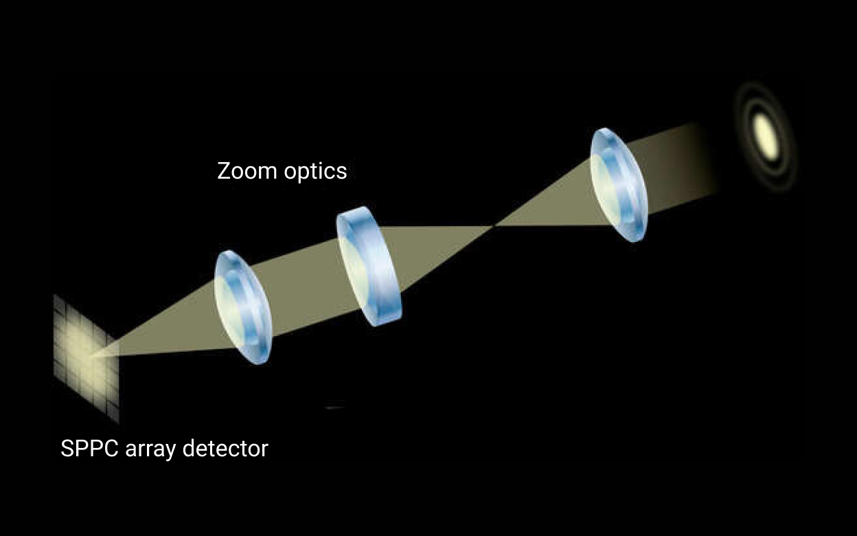
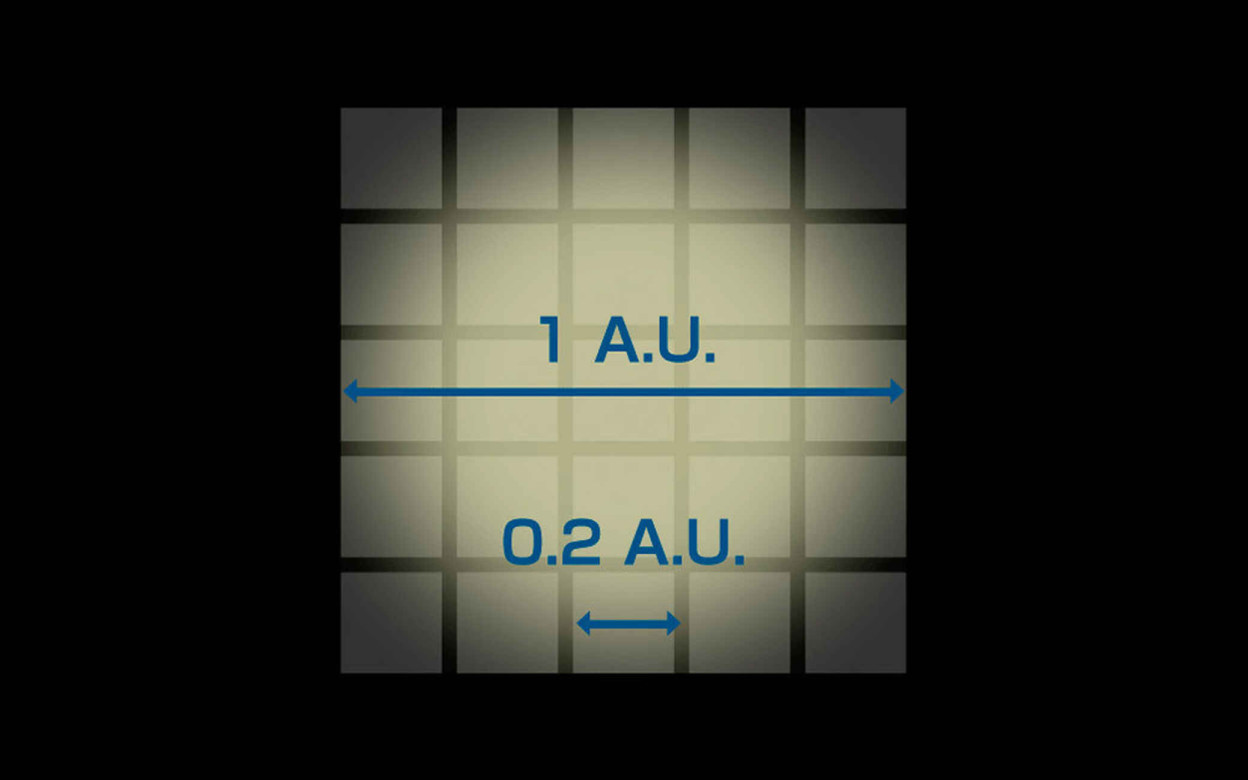
Since light is a wave and has the property of diffraction, when a single point of light is imaged, the image exhibits a certain degree of spread. This spread makes it impossible to distinguish between two points of light that are close together, which is called the theoretical limit of resolution. In microscopy, by modifying illumination, observation targets, and detection methods, it is possible to capture fine structures beyond the theoretical limit of resolution. As explained below, there are ways to improve lighting by using striped illumination and image reconstruction to achieve super-resolution. In designing observation targets, there is a method for fluorescence observation that separates and detects adjacent fluorescence by utilizing the spatial distribution of fluorescence blinking over time. For detection, there is a method of detecting light from an imaging area smaller than the laser beam irradiation spot when observing the optical response of a target to laser irradiation.
Click to enlarge
Nikon provides products featuring various super-resolution microscopy techniques. In fluorescence imaging with STORM (Stochastic Optical Reconstruction Microscopy), fluorescent molecules are excited, and the signals are measured by distinguishing neighboring fluorescent molecules by time differences. In SIM (Structured Illumination Microscopy), the target object is illuminated with patterned illumination, and information on the microstructure is obtained using the moiré effect. Simultaneously, these microscopy techniques, including confocal microscopy, need to observe samples quickly, avoid fluorescence photobleaching, and reduce noise from out-of-focus regions in thick samples.
Technology Application Examples
Structured Illumination Microscope
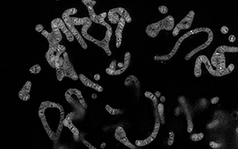
The Super Resolution Microscope N-SIM series attains spectacularly high resolution of nearly double that of conventional optical microscopes, by combining Nikon's unique optical technologies and Structured Illumination Microscopy (SIM), a technique announced by Dr. Mats G.L. Gustafsson et al. in the University of California, San Francisco in 2000.
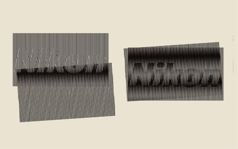
When a finely striped, patterned illumination (structured illumination) is applied to a specimen, it produces moiré patterns. These patterns are characteristically coarser than the original and can be captured by optical microscopes, even when observing the finer structures of the specimen.
N-SIM captures multiple moiré images while slightly varying the orientation and phase of the structured illumination. Then it analytically processes those images to mathematically recreate the intricate structures of the specimen.
N-SIM achieves significant resolution by using a new illumination scheme without drastically altering the microscope mechanism.
Additionally, Nikon has developed an innovative high-speed, structured illumination system. The N-SIM S Super Resolution Microscope realizes high-speed, super-resolution imaging.

Technologies related to these examples
Related Technology
Microscopy
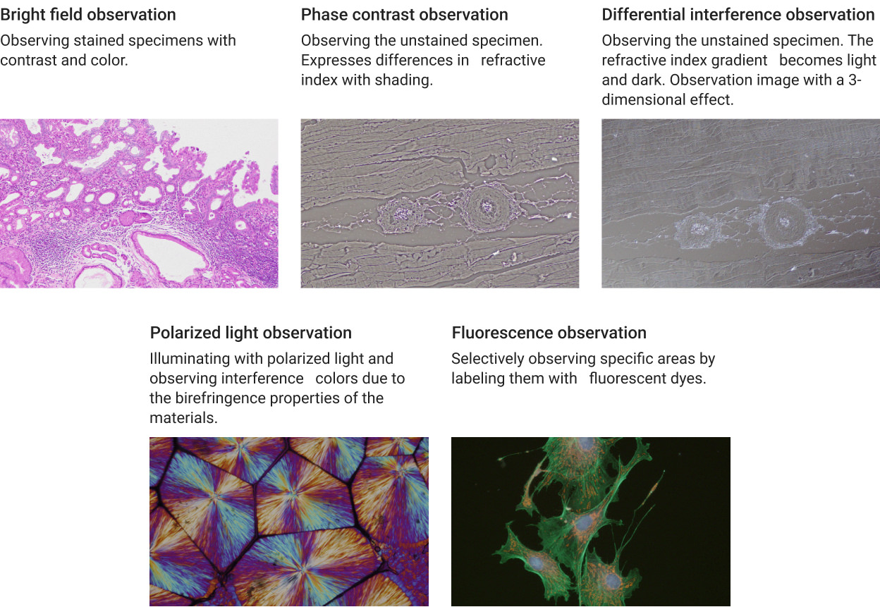
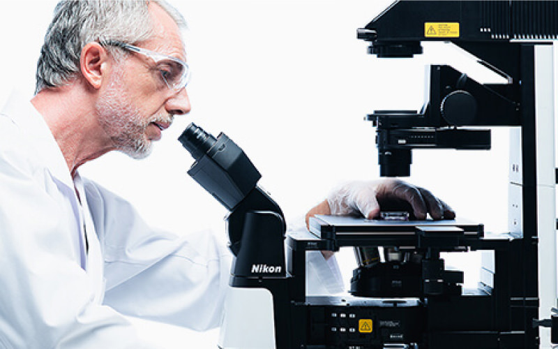
Microscopy is a technology that makes small objects visible and is used not only for observing living organisms such as cells but also for industrial applications, such as observing minerals and measuring electronic components. By capturing and analyzing observed images as data, it is used not only for observation but also for measurement.
Furthermore, microscopy is used not only for academic discoveries in biology, but also for measurements and examinations in the biotechnology industry, such as diagnosis in medical settings and drug discovery.
At Nikon, we use microscopy to support a variety of applications, such as the biological observation of cells and inspection of semiconductor wafers, electronic components, and materials. In biological and clinical/biomedical research, there is a demand for quantitative observation of larger living cells in real time, which requires high-speed, high-resolution observation methods. For high-resolution observation, it is important to scan each point at high speed while ensuring that the necessary signal is sufficiently obtained against noise.
Main Related Products
You can search for articles related to Nikon’s technology, research and development by tag.

