Nikon introduces the AX R MP with NSPARC Super-resolution Multiphoton Confocal Microscope
Using super resolution to observe deep areas within living organisms and expand our understanding of brain diseases
July 27, 2023

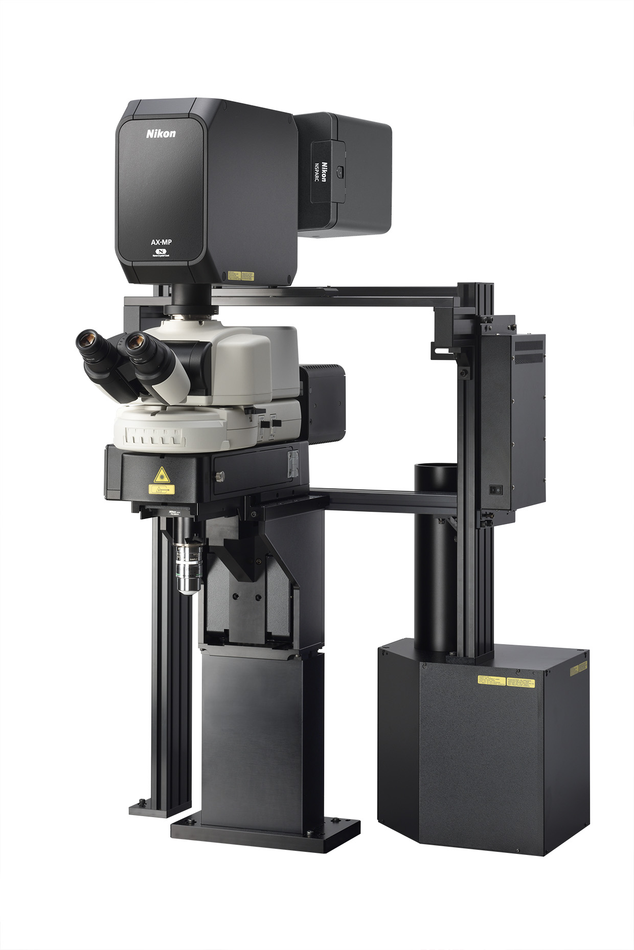
(configured with AX-FNSP microscope)
TOKYO - Nikon Corporation (Nikon) has announced the release of the AX R MP with NSPARC super-resolution multiphoton confocal microscope, which enables extremely sensitive array detection and super-resolution in deep areas within large living organism specimens. This product will contribute to the understanding of brain diseases such as Alzheimer's and Parkinson's disease, as well as drug discovery research.
Release Overview
Swipe horizontally to view full table.
| Product Name | AX R MP with NSPARC super-resolution multiphoton confocal microscope |
|---|---|
| Release Date | Beginning of September, 2023 |
Development Background
In recent years, multiphoton microscopy has been used to drive scientific understanding of topics such as real-time neuronal activity and drug efficacy in living organisms.
Nikon has been in a leader in super-resolution microscopy since 2010, offering many technologies that exceed the resolution limit of general optical microscopes. In addition, Nikon released the AX R MP multiphoton confocal microscope last year, providing high-resolution, high-speed, large field-of-view images in deep areas within living bodies.
Now, by combining the unmatched sensitivity of array-based NSPARC detection and the optical superiority of the AX R MP, super-resolution observation of deep areas within living bodies can be realized. The combination of these technologies will contribute to nervous system drug discovery research helping to advance knowledge around diseases such as Alzheimer's, Parkinson's and mechanisms of dementia.
Main Features
1. Observe fine structures deep within living bodies by combining multiphoton microscopy with super-resolution.
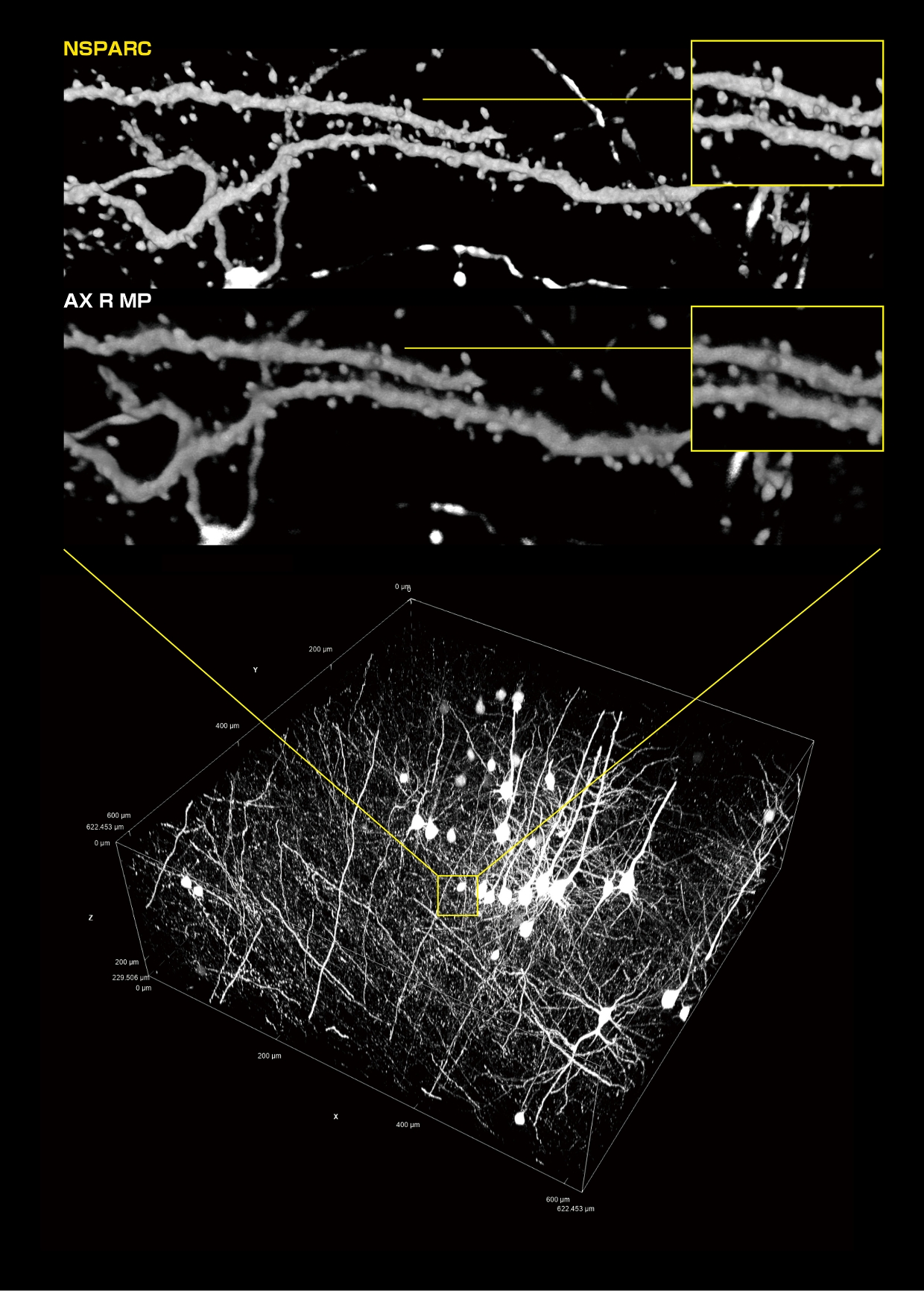
With the addition of NSPARC-based detection to the AX R MP, low-noise super-resolution detection can be applied to image deep within large, living specimens. In addition, combining low-noise array detection with a high-speed resonant scanner allows researchers to get a clear picture of the rapid dynamics within the living brain. These images can drive analyses that may link neuronal disease mechanisms with neuronal morphology and dynamics. Visualizing the fine structures of the central nervous system using this combination can bring forth more information than normal detectors, contributing to the understanding of their functions.

(within yellow frames: identifying the shape and size of a spine neck can be expected to contribute to the clarification of neural activity)
Sample courtesy of: Lin Daniel, PhD., SunJin Lab Co.
2. Utilize Nikon-developed AI to increase the efficiency of imaging and analysis workflows.
With a rapidly expanding suite of AI tools, super-resolution images acquired with AX R MP with NSPARC can be efficiently captured, processed and analyzed using Nikon's imaging software, NIS-Elements. AI automatically performs complex image processing after imaging, such as noise removal and target cell extraction. These powerful AI functions can reduce user workloads, improve data collection and enhance image analysis.
(“such as brightness adjustment, noise removal,” was changed to “such as noise removal” on August 9, 2023.)

(The Autosignal.ai image and caption were deleted on August 9, 2023.)
3. World-class objectives suitable for observation of deep parts of brain tissue
By controlling the production of optics from the glass itself, Nikon provides a wide range of class-leading high-resolution objectives for multiphoton microscopy. These lenses can be used in the wavelength range from ultraviolet to near-infrared with minimal color bleeding, enabling selection of a lens according to the purpose of a particular experiment. The CFI Plan Apochromat Lambda S 60XC Sil, which is suitable for super-resolution deep observation at high magnification, has also been newly added to the lineup, which can contribute to further nervous system mechanisms clarification. As the NIS-Elements control software and objective information are linked, image analyses can be performed without the need for complicated parameter settings.

At the left end is the CFI Plan Apochromat Lambda S 60XC Sil released on May 17
Near-IR Imaging Option expands the multiplexing capabilities of Nikon's AX and AX R confocal microscopes.
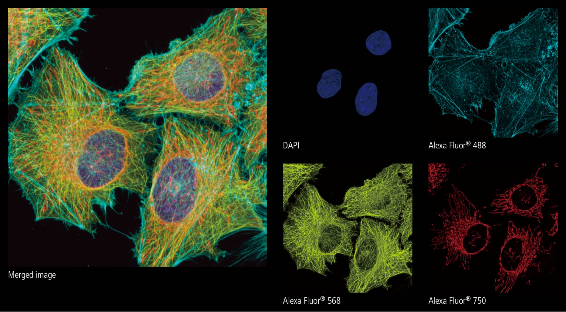
Further demonstrating Nikon's commitment to continued development of current product offerings, the NIR Imaging Option, for use in combination with the AX and AX R confocal microscopes, will also be released simultaneously with the AX R MP with NSPARC. This option adds specialized NIR-sensitive excitation and detection technology to AX confocal systems. This enables the addition of near-infrared probes to existing visible light excitation, broadening the possibilities of research.
With the AX / AX R confocal microscope and AX R MP with NSPARC, Nikon continues to make products available to support research on life phenomena, and expand its range of products, providing the flexibility to meet customers' research needs.
Swipe horizontally to view full table.
| Product Name | NIR Imaging Option near-infrared observation unit for AX / AX R confocal microscope |
|---|---|
| Release Date | Beginning of September, 2023 |

Nucleus (DAPI), Actin (Alexa Fluor® 488), Tubulin (Alexa Fluor® 568),
Mitochondria (Alexa Fluor® 750)
The information is current as of the date of publication. It is subject to change without notice.
