Nikon introduces the new Research Stereo Microscopes SMZ25/SMZ18
May 13, 2013
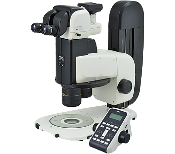
Nikon Corporation (Makoto Kimura, President; Chiyoda-Ku, Tokyo,) is pleased to announce the introduction of our newest Research Stereo Microscope SMZ25/SMZ18, featuring "the largest zoom range"*1, "extremely high resolution" and "exceptionally bright Epi-fluorescence".
The SMZ25/SMZ18 is ideally suited for both bioscience and medical fields as well as industrial fields. The extremely large zoom range and high resolution allow users to image the sample from its entirety down to its microscopic details.
- *1With SMZ25 (as of May 2013).
Product information
Product name : Research Stereo Microscope SMZ25 / SMZ18
Available from June 3, 2013

Motorized Epi Fluorescence set

Episcopic Plain Stand set
Background of Development
Biomedical fields including molecular biology, cell biology, neurobiology, embryology, developmental biology and systems biology have increasing needs for imaging systems that span spatial scales from single cells to whole organisms, whilst maintaining the wide field of view, highest resolution and fluorescence transmission. With these demands in mind, Nikon has developed two new research stereo microscopes, the motorized SMZ25 and manual SMZ18. Both feature extremely high resolutions and exceptional fluorescence transmission capabilities. SMZ25 also features the largest zoom range on the market*2.
- *2As of May 2013
Key features
1. The largest zoom range*3 (25:1) and exceptional resolution
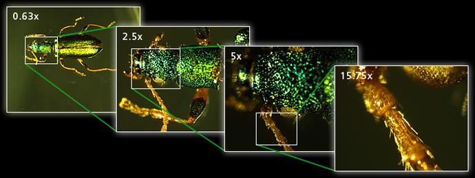
The new Nikon stereo microscopes incorporate a newly developed zoom system (Perfect Zoom System). The Perfect Zoom System is a breakthrough in stereo microscope design, enabling both an incredible zoom range of 25:1 (0.63-15.17x) and a high numerical aperture of 0.156 (for SMZ25, 1x Objective, at the highest zoom), which allow researchers to image both the entire sample as well as its microscopic details using just a single instrument.
Especially, the SMZ25 boasts a 35mm field of view at the lowest magnification of 0.63x (using 1x objective, 10 x eyepieces), enabling users to image the entire 35mm dish. At the high magnification range, Nikon's newly developed high performance objective lenses allow imaging of microscopic structures once considered too small to visualize on a stereo microscope.
- *3With SMZ25 (as of May 2013).

Using the 1x objective lens with SMZ25
Image courtesy of Japan Insect Association
2. A breakthrough in Epi-fluorescence imaging: stunningly bright images and high contrast
The new epi-fluorescence attachment employs a fly-eye-lens that enables brighter and more uniform illumination across the entire field of view even at the low magnification range. In addition, the signal-to-noise ratio has been improved by the incorporation of a short wavelength, high transmission lens in the zoom body. The result is clear, high contrast fluorescent imaging.
3. Wide range of accessories
A wide variety of accessories such as illuminators, bases and stands are available to accommodate the wide-range of research needs in the biomedical and industrial fields. Nikon's imaging software, NIS-Elements (sell separately), can be now be used to operate SMZ25 to capture multi-channel, time-lapse images, and Extended Depth of Focus (EDF)*4 images. By combining the SMZ25 or SMZ18 with Nikon's stand alone control unit DS-L3 (sell separately), researchers can easily visualize critical information such as magnification and diascopic illumination intensity on the LCD monitor and also configure camera acquisition settings using the touch panel.
- *4A single extended depth of focus image created from a stack of images taken at different focal depths
4. More ease of use
The thinner LED diascopic illumination base increases the efficiency and ergonomics of sample manipulation and exchanging of samples. The new base also features a built-in Nikon's unique OCC (Oblique Coherent Contrast)*5 illuminator that produces high-contrast images of transparent samples such as ITO film and zebrafish. The SMZ25 also boasts an easy-to-use remote controller. The new remote controller with focusing knob enables the researcher to change focus, zoom, and intensity of the diascopic LED light with ease, and can also be used to capture images.
- *5The OCC stands for Oblique Coherent Contrast. The OCC method uses the slide diaphragm located near the position of the entrance pupil of an objective to shield luminous flux and applies coherent light to samples diagonally. This gives shades to colorless transparent samples so they can be observed with contrast.
Specification
Swipe horizontally to view full table.
| SMZ25 | SMZ18 | ||
|---|---|---|---|
| Total magnification (Using 10x eyepiece) |
3.15x-315x (Depending on objective used) |
3.75x-270x (Depending on objective used) |
|
| Zooming Body | Optical system | Parallel-optics (zooming type) | Parallel-optics (zooming type) |
| Zoom range | 0.63-15.75x | 0.75—13.5x | |
| Zoom ratio | 25:1 | 18:1 | |
| Zoom | Motorized | Manual | |
| Aperture diaphragm | Zooming body built-in | Zooming body built-in | |
| Controller | Remote controller | — | |
| Objectives NA / Working Distance (mm) |
0.5x | 0.078 / 71 | 0.075 / 71 |
| 1x | 0.156 / 60 | 0.15 / 60 | |
| 1.6x | 0.25 / 30 | 0.24 / 30 | |
| 2x | 0.312 / 20 | 0.3 / 20 | |
| Tubes | Tilting trinocular tube / Low eyelevel trinocular tube | ||
| Epi-Fluorescence Attachment | Motorized / Manual filter cube turret (4 filter cubes mountable) | ||
| Bases / Stand | LED diascopic base / Fiber diascope base Episcopic plain base / Episcopic plain stand |
||
| Episcopic illuminators | Coaxial Epi illuminator / LED ring illumination unit LED ring fiber illumination unit Flexible double arm fiber illumination unit |
||
| Diascopic illuminators | LED dark field unit / Polarizing attachment | ||
The information is current as of the date of publication. It is subject to change without notice.
